Скачай курс
в приложении
Перейти в приложение
Открыть мобильную версию сайта
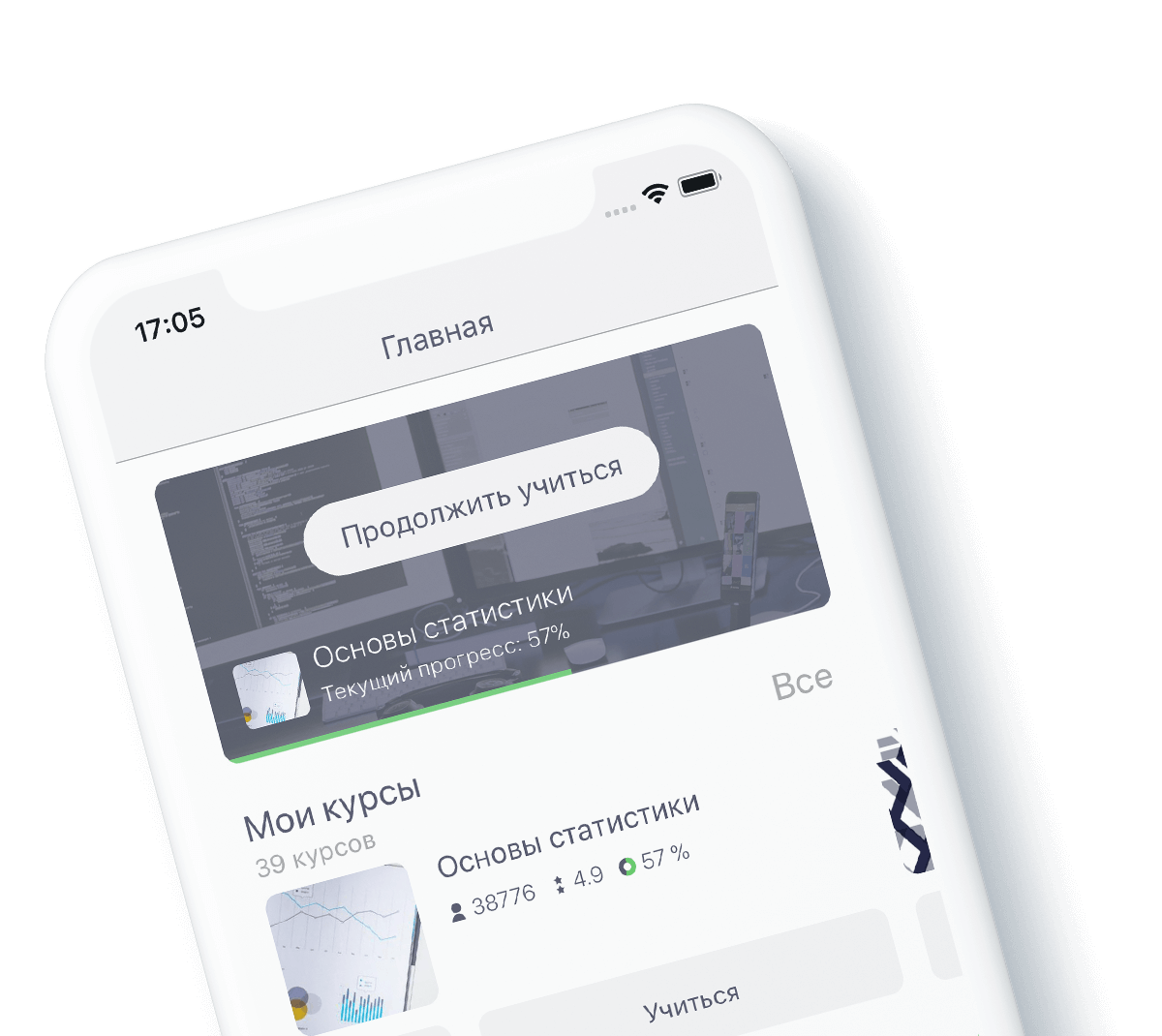
Каталог
Моё обучение
загрузка...